Chondrodermatitis Nodularis Helicis
Medically reviewed by Dennis A Porto, MD and The Dermatologists
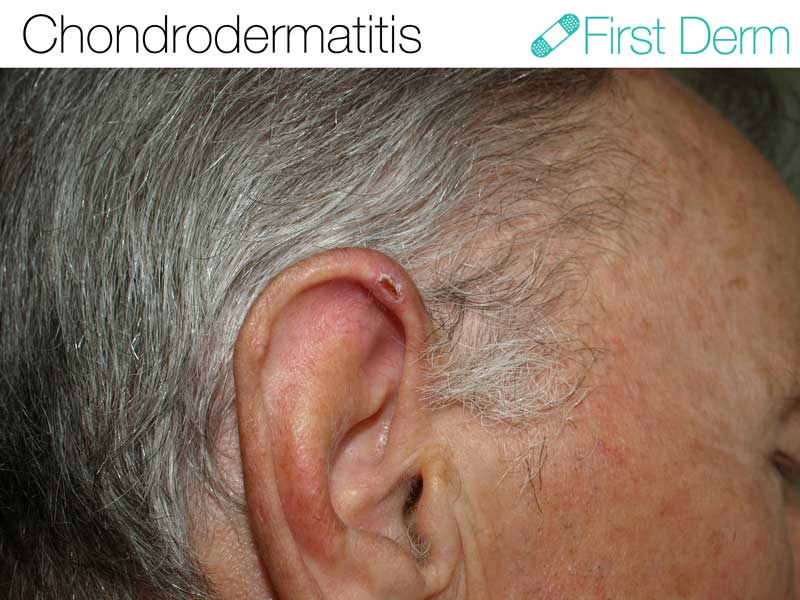
![Chondrodermatitis nodularis helicis (01) örat [ICD-10 H61.039]](https://www.firstderm.com/wp-content/uploads/Chondrodermatitis-nodularis-helicis-01-örat-ICD-10-H61.039.jpg)
Chondrodermatitis nodularis helicis (01) örat [ICD-10 H61.039]
Chondrodermatitis-Nodularis-Chronica-Helicis-ICD-10-H61
More than 200,000 US cases per year
- Requires medical diagnosis
- Symptoms: Painful, firm bump covered by crust
- Color: Typically red-brown
- Location: Outer part of the ear
- Treatment: Topical steroids, excision, cortisone injection
ICD-10: H61.039
ICD-9: 380.03
Chondrodermatitis nodularis helicis is a lesion on the helix or antihelix (the raised portions) of the ear. The condition is more common in men over the age of 40. Only 10-35% of cases involve women.
Although the exact cause is unknown, it occurs most often in people who tend to sleep on one side. Exposure to cold, a telephone headset, or tight headgear can also trigger chondrodermatitis.
Chondrodermatitis nodularis helicis occurs in people of all races, but most commonly in fair-skinned individuals with sun-damaged skin. It may occasionally be associated with autoimmune or connective-tissue disorders.
Try our FREE dermatology search engine and get peace of mind within a second
Symptoms
Chondrodermatitis nodularis helicis is typically painful and crusty. It is an inflammatory condition that manifests itself in a solitary firm scaly bump.
A lesion can grow to 2-4mm in diameter in a few months and remain the same size. It may also excrete a scaly discharge. The condition can last for months or years without treatment.
The condition is localized, but can cause discomfort when pressed or when it is cold. The pain can be intense but is usually short-lived, though it can last for as long as an hour.
What can I do?
Treating involves eliminating pressure on the ear. Avoid sleeping on the side with the affected ear, and protect your ears with a hat when outside in the cold. You can also try to alter the way you hold your phone to the ear or put on a corn plaster. Make sure your pillow is soft, and you can make a hole at the area that comes in contact with the bump.
Should I seek medical care?
Although these nodules are benign, they are are similar in appearance to various skin cancers.
Therefore, you should exercise caution with these lesions and see a doctor in order to rule out the possibility of a skin malignancy. A biopsy is occasionally needed to differentiate the lesion from skin cancer forms, such as basal cell carcinoma and squamous cell carcinoma.
Treatment
There are several effective treatment options. Topical or steroids or steroid injections can help. Freezing with liquid nitrogen by a dermatologist has also been used with success. Excision is not typically the first choice, but is another option. Lesions can recur, unfortunately, which can be frustrating to patients and dermatologists alike.
Try our FREE dermatology search engine and get peace of mind within a second
Source:
British Association of Dermatologists. CHONDRODERMATITIS NODULARIS. Available at: https://www.bad.org.uk/shared/get-file.ashx?id=75&itemtype=document
Primary Care Dermatology Society. Chondrodermatitis Nodularis Helicis. Available at: https://www.pcds.org.uk/clinical-guidance/chondermatitis-nodularis-helicis
Image courtesy of Klaus D. Peter, Gummersbach, Germany via Wikicommons, First Derm watermark added.
Ask a Dermatologist
Anonymous, fast and secure!

The Specialist doctor from the University Hospital in Gothenburg, alumnus UC Berkeley. My doctoral dissertation is about Digital Health and I have published 5 scientific articles in teledermatology and artificial intelligence and others.