Nodular melanoma
Medically reviewed by The Dermatologists and written by Dr. Carol Mastropierro and Dr. Alexander Börve
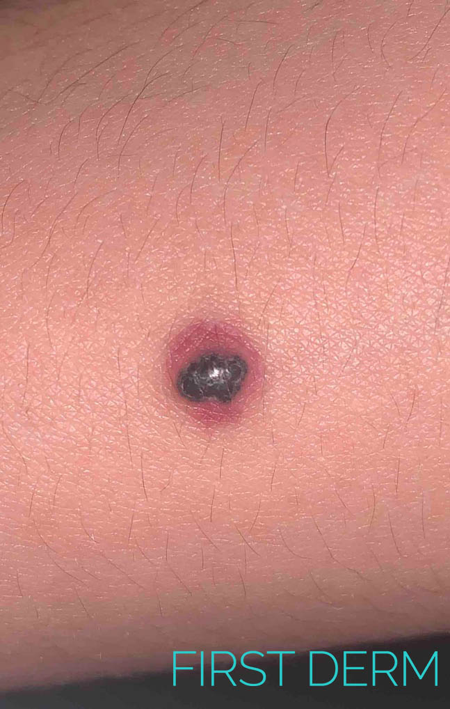
Nodular melanoma (1) skin

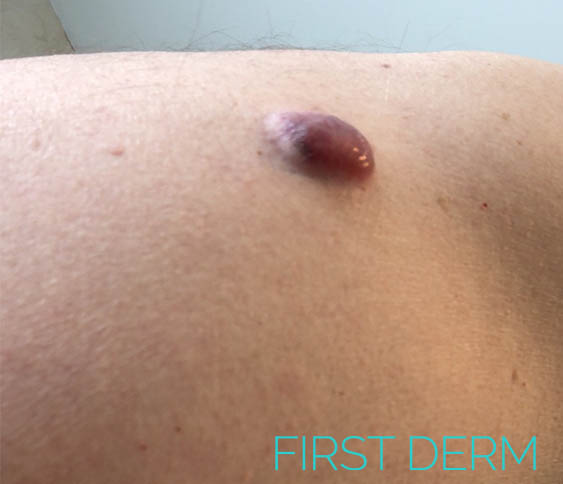
Nodular melanoma (2) skin
Nodular melanoma (3) skin
An example of Nodular Melanoma.
Common
- Nodular melanoma accounts for 20-25% of cases of melanoma and is the most aggressive type.
- Usually appear as a changing lump on the skin that might be black to red in colour.
- Most commonly grow on the head and neck, chest or back.
- Bleeding or oozing is a common symptom.
- Diagnosis is by dermoscopy and skin biopsy. Lymph node biopsy is advised for lesions more than 1mm thickness.
- Treatment by complete excision with a 2-3 cm margin of normal tissue
Melanoma is a potentially dangerous skin cancer that arises from pigment cells known as melanocytes. Nodular melanoma is an invasive form of melanoma which accounts for roughly one fourth of diagnosed melanomas. It is characterized by being faster developing, meaning it quickly grows downwards into the deeper layers of skin.
Nodular melanoma has higher incidence in males over the age of 50 than in females. Although it is more common in fair skin phototypes, it may also occur in those who tan quite easily (phototype 3), and occasionally in brown or black skin (phototype 4-6).
Consult an online dermatologist today and get an answer on your skin concern within hours.
Symptoms
Nodular melanomas usually appear as a changing lump or nodule on the skin that might be black to red in color. However, one-third of nodular melanomas are not pigmented and lack the ABCDE criteria (Asymmetry, Border irregularity, Color variation, Large Diameter).
They often grow on previously normal skin and most commonly grow on the head and neck, chest or back. It is less strongly associated with sun exposure than superficial spreading and lentigo maligna types of melanoma.
Bleeding or oozing is a common symptom, but not always present.
Consult an online dermatologist today and get an answer on your skin concern within hours.
What can I do?
It is advisable for you to keep an eye out for any change in the appearance of your skin. Any newly arising nodular lesion or nodule from a preexisting mole should be addressed to exclude melanoma. Although you can self-examine your skin following the ABCD rule, it is important for you to bring your mole to the attention of a dermatologist.
You should attend routine check ups with your doctor, especially if you have risk factors, such as older age, previous invasive melanoma or melanoma in situ. It should be noted that you are also high risk if you have a family history of skin cancer or fair skin that burns easily.
Consult an online dermatologist today and get an answer on your skin concern within hours.
Should I seek medical care?
If you notice a funny- looking mole or a change in an existing mole you should make an appointment with your dermatologist as soon as possible. Your doctor will look at the lesion through a dermoscope, a special lens that magnifies it. Signs of asymmetry, atypical vascular pattern and multiple colours are signs of malignancy. Your doctor will definitely take a skin biopsy, usually an excision biopsy and send a sample to a pathologist.
The prognosis and treatment plan will be given after the pathologist determines the Braslow’s score. This measures the extension of the melanoma into the dermis. The thicker the melanoma, the more likely it is to metastasize (spread to distant sites). For this reason, melanomas that are more than 1 mm thick may need to have lymph node biopsy as well.
Consult an online dermatologist today and get an answer on your skin concern within hours.
Treatment
Initial treatment of primary melanoma is by resection. The lesion should be completely excised with a 2-3 cm margin of normal tissue. Further treatment depends mainly on the Breslow thickness of the lesion. This is decided after the resected tumor is analysed by a pathologist under a microscope. In the event the pathologist determines that the melanoma was excised with incomplete margins, adjuvant radiotherapy is recommended.
If regional or distal lymph nodes are enlarged, suspicion for lymph nodes involvement is raised. A test called sentinel node biopsy is run. If metastases are found, the affected lymph nodes must be surgically removed.
Consult an online dermatologist today and get an answer on your skin concern within hours.
Source:
Erkurt MA, Aydogdu I, Kuku I, Kaya E, Basaran Y. Nodular melanoma presenting with rapid progression and widespread metastases: a case report. J Med Case Rep. 2009;3:50. Published 2009 Feb 6. doi:10.1186/1752-1947-3-50: https://www.ncbi.nlm.nih.gov/pmc/articles/PMC2644319/
Ask a Dermatologist
Anonymous, fast and secure!

The Specialist doctor from the University Hospital in Gothenburg, alumnus UC Berkeley. My doctoral dissertation is about Digital Health and I have published 5 scientific articles in teledermatology and artificial intelligence and others.