Melanoma Pictures and Malignant Tumor of The Skin
Malignant Melanoma Pictures
Malignant melanoma is the most serious form of skin cancer and the cancer form that increases the most among humans. It is strongly related to sun UV rays and repeated sun-burns. If detected early and treated early, 95% survive the disease. Therefore early detection is important.
The disease is very rare before puberty and unusual during adolescence. Healthy sun habits are important during childhood, which reduces the risk of getting malignant melanoma when you get older. Today we’ll take a look at some malignant melanoma pictures so that you can spot the signs early.
Symptoms
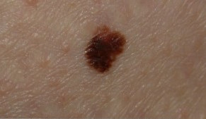
Malignant melanoma is most common on the chest, back and legs, but can occur anywhere on the skin. The most common symptoms is a new brown-black spot that has grown or changed appearance. It may also look like a birthmark or a mole that begins to change. Malignant melanoma is often:
- Irregular in shape and uneven at the edge
- Uneven in color: for example, different shades of brown, black, red, pink, blue or white.
- More than five millimeters.
- Moles that bleed can also be signs of the disease.
Common birthmarks, called nevi, are usually even brown in color and there is almost always a regular limit to the skin around. Wart like and hairy moles are rarely signs of cancer.
By taking a look at our skin guide you can learn more about malignant melanoma as well as more melanoma pictures.
There are also other types of skin cancer. The most common form is basal cell carcinoma, followed by squamous cell carcinoma.
Try our FREE dermatology search engine and get peace of mind within a second.
Identifying Malignant Melanoma: Real Life Cases
[Consulted by First Derm Dermatologists]
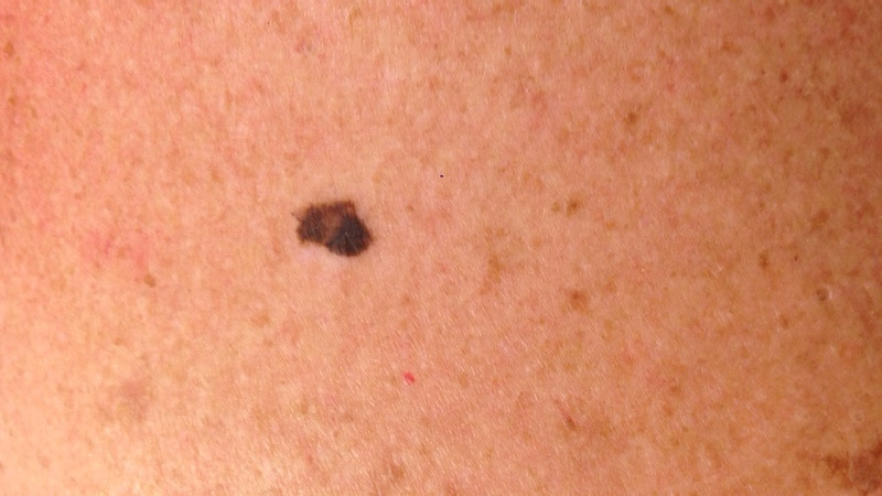
Case 1: Dark Mole On Chest
Online dermatology question
I am a 40 year old female. Dark mole on my chest, present for a long time but has recently become bigger and more irregular. Width 6mm.
Online dermatology answer
Thank you for sending your case. Based on the information and images, this is possibly a MALIGNANT MELANOMA. Melanoma is a type of skin cancer usually caused by excessive sun exposure, especially a history of sunburn(s). The pigmented lesion seen in the images is larger than most benign moles and also shows an irregular shape and multiple colors.
An urgent visit (within 2 weeks) to a see a dermatologist for closer examination with dermoscopy (a diagnostic technique with a specialized magnifying glass which allows for a more precise assessment) followed by urgent surgical excision is highly recommended.
Try our FREE dermatology search engine and get peace of mind within a second.
Case 2: Dark Mole On Back
Online dermatology question
I am a 65 year old male. Dark mole left side of back. No symptoms. Wife concerned that it might have gotten bigger.
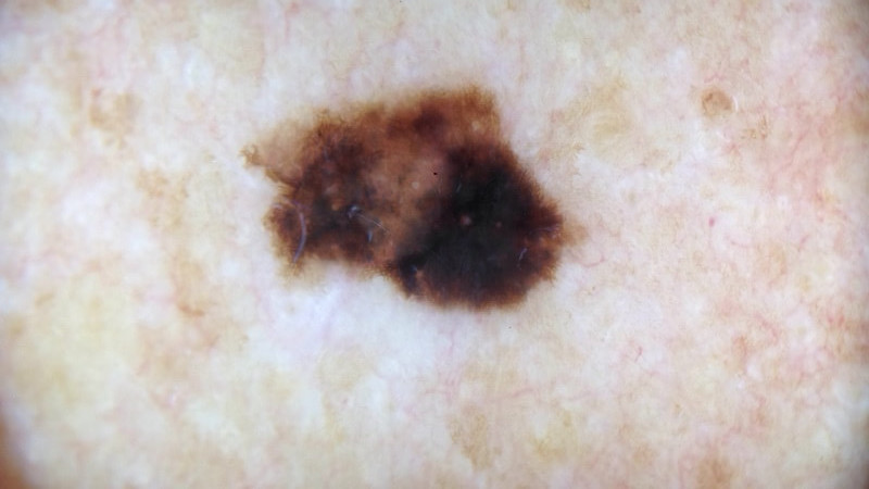
Online dermatology answer
Based on the information and images, this is possibly a MALIGNANT MELANOMA: Melanoma is a type of skin cancer usually caused by excessive sun exposure, especially a history of sunburn(s). The pigmented lesion seen in the images is larger than most benign moles and also shows an irregular shape and multiple colors.
Nevertheless, it is hard to be sure. Melanoma pictures can be distorted and this could also be a benign mole known as seborrhoeic keratosis (benign wart). In any case, an urgent visit to a see a dermatologist for closer examination. I recommend that you store these images with this answer and bring them with you.
Try our FREE dermatology search engine and get peace of mind within a second.
Case 3: Lighter Lesion On Face
Online dermatology question
I am a 44 year old male. It is on my face. Lighter lesion newer and growing. I am a farmer and I have been in the sun a lot. I removed a superficial spreading melanoma en situ last week from my back
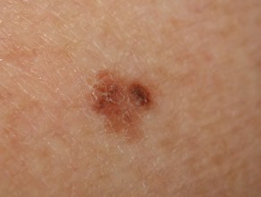
Online dermatology answer
Based on the information and images, this is possibly a MALIGNANT MELANOMA: Melanoma is a type of skin cancer usually caused by excessive sun exposure, especially a history of sunburn(s). The pigmented lesion seen in the images is larger than most benign moles and also shows an irregular shape and multiple colors.
As mentioned with previous melanoma pictures, it could also be a benign so-called seborrhoeic keratosis (benign wart). In any case, an urgent visit to a see a dermatologist for closer examination with dermoscopy (a diagnostic technique with a specialized magnifying glass which allows for a more precise diagnosis) is highly recommended. At this visit the other lesion should also be assessed since this also can be a skin cancer since it’s new and growing.
Try our FREE dermatology search engine and get peace of mind within a second.
Case 4: Dark red slightly raised in center surrounded by a light red color – Leg
Online dermatology question
I am a 49 year old woman. Location is right back of leg, dark red slightly raised in center surrounded by a light red color. The dark red part of the mole is 1/2 cm and 1 cm if you include the light red part. Not sure if the light red area is actually part of the mole or did I irritate the mole while wearing a pair of jeans and the light red area is a bruise around the mole.
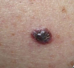
Online dermatology answer
Based on the information and images, this is possibly a MALIGNANT MELANOMA: Melanoma is a type of skin cancer usually caused by excessive sun exposure, especially a history of sunburn(s). The pigmented lesion seen in the images is larger and darker than most benign moles. It shows a dark blue-red and black and colors with bleeding surrounding it.
Nevertheless, it is hard to be sure. It could also be a benign mole that is very dark and bleeding but it is impossible to exclude without inspecting it with a dermoscope. In any case, an urgent visit to a see a dermatologist for closer examination with dermoscopy (a diagnostic technique with a specialized magnifying glass which allows for a more precise diagnosis) s highly recommended.
Try our FREE dermatology search engine and get peace of mind within a second.
Sources:
U.S. Cancer Statistics Working Group. United States Cancer Statistics: 1999–2012 Incidence and Mortality Web-based Report. Atlanta: U.S. Department of Health and Human Services, Centers for Disease Control and Prevention and National Cancer Institute; 2015. Available at: https://www.cdc.gov/cancer/dcpc/data/
AIM at Melanoma Foundation. Melanoma Stats, Facts, and Figures. Available at: https://www.aimatmelanoma.org/about-melanoma/melanoma-stats-facts-and-figures/.
American Academy of Dermatology. Melanoma. Available at: https://www.aad.org/public/diseases/skin-cancer/melanoma
National Cancer Institute. SEER Stat Fact Sheets: Melanoma of the Skin. Available at: http://seer.cancer.gov/statfacts/html/melan.html
Ask a Dermatologist
Anonymous, fast and secure!
Over 15,000+ Readers
Get fresh content from First Derm
![]()
![]()
![]()
Ask a Dermatologist
Anonymous, fast and secure

The Specialist doctor from the University Hospital in Gothenburg, alumnus UC Berkeley. My doctoral dissertation is about Digital Health and I have published 5 scientific articles in teledermatology and artificial intelligence and others.